Dividing cancer cell, fluorescent microscopy - HD Stock-Video
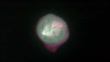
Timelapse fluorescence microscopy video of a dividing cancer cell in 3D tissue culture. The total length of the sequence is 3 hours. The cell is expressing a fluorescently-tagged version of a protein (EB1) that binds to growing microtubule ends and also localizes to the spindle poles. This highlights the highly dynamic microtubule cytoskeleton in the spindle, and also shows astral microtubules extending toward the cell periphery. The different colors represent different focal planes giving a more 3D impression. In the beginning of the video it can be seen how the duplicated centrosomes move around the cell nucleus and then form the poles on opposite ends of the mitotic spindle apparatus. Although chromosomes themselves are not labeled, they can be seen as darker shadows in the cytoplasm. The cell eventually divides apparently without all chromosomes having aligned correctly in the middle of the spindle, leading to aneuploidy, one of the hallmarks of cancer. A slowed down version of this clip can be seen as K007/4486


EINE LIZENZ KAUFEN
Individuelle Preisgestaltung: Sagen Sie uns einfach, wann, wo und wie Sie diese Datei nutzen möchten.
DETAILS
Bildnachweis:
Creative #:
1432329434
Lizenztyp:
Rights-ready
Kollektion:
Photolibrary Video
Max. Dateigröße:
1920 x 1080 px - 271 MB
Cliplänge:
00:00:21:16
Hochgeladen am:
Releaseangaben:
Keine Freigaben erforderlich
Gemastert mit:
QuickTime 8-bit Photo-JPEG HD 1920x1080 25p
Kategorien:
- Krebszelle,
- Krebs - Tumor,
- Handy,
- Biologie,
- Onkologie,
- Bizarr,
- Wiederholung,
- Vergrößerung,
- Zeitraffer,
- Chromosom,
- Mikrotubulus,
- Forschung,
- Metastatischer Tumor,
- Schwarzer Hintergrund,
- Zellteilung,
- Anaphase,
- Cytoskeleton,
- Drahtlose Technologie,
- Farbbild,
- Film - Filmtechnik,
- HD-Format,
- Hohe Aufnahmegeschwindigkeit,
- Horizontal,
- Karyokinese,
- Krankheit,
- Lichtmikroskopische Aufnahme,
- Metaphase,
- Mitotisch,
- Prophase,
- Spindel,
- Wissenschaftliches Experiment,
- Zustand,